

Introduction to Oral Tolerance and Immune Regulation
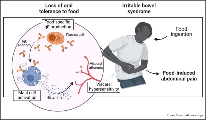
Oral tolerance is a fundamental immunological process in which exposure to food antigens through the gastrointestinal tract suppresses subsequent systemic immune responses against the same antigens. This phenomenon has been extensively studied in rodents and is also observed in humans and other species.
The intestinal immune system continuously encounters large amounts of dietary proteins and microbial antigens. Despite this constant antigen exposure, harmful immune reactions against food components are normally prevented. This controlled immune suppression is essential for maintaining intestinal homeostasis and preventing food allergies or chronic inflammation.
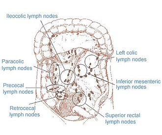
Recent studies demonstrate that mesenteric lymph nodes (MLNs) play a central role in the induction of oral tolerance by coordinating communication between the intestinal mucosa and the systemic immune system.
Food Antigens and Systemic Immune Tolerance
During digestion, some food proteins and protein fragments cross the intestinal barrier and enter the lymphatic and blood circulation. These antigens are transported to secondary lymphoid tissues where immune regulation occurs.
Oral tolerance suppresses both:
- Cellular immune responses
- Humoral immune responses including IgE, IgG, and IgM production
This systemic hyporesponsiveness depends mainly on CD4+ T lymphocytes.
Experimental studies show that:
- Removal of CD4+ T cells abolishes oral tolerance
- Transfer of tolerant CD4+ T cells can induce tolerance in naïve animals
- Tolerized T cells fail to provide effective B-cell activation signals
As a result, antibody-producing B cells become functionally suppressed.
Mechanisms of T Cell Hyporesponsiveness
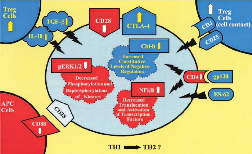
The immunological mechanisms involved in oral tolerance are complex and influenced by antigen dose, exposure conditions, and immune status.
Several major mechanisms contribute to T-cell suppression:
1. Clonal Deletion
High antigen concentrations can eliminate antigen-specific T cells through apoptosis.
2. T Cell Anergy
Lower antigen doses may induce functional inactivation of T cells without cell death.
3. Regulatory T Cells (Tregs)
Regulatory T cells actively suppress immune responses through anti-inflammatory cytokines such as TGF-β.
Important Treg populations involved include:
- CD4+ TGF-β-producing T helper 3 cells
- CD4+ CD25+ FoxP3+ regulatory T cells
These cells contribute to long-term immune tolerance toward food antigens.
Role of Dendritic Cells in Oral Tolerance
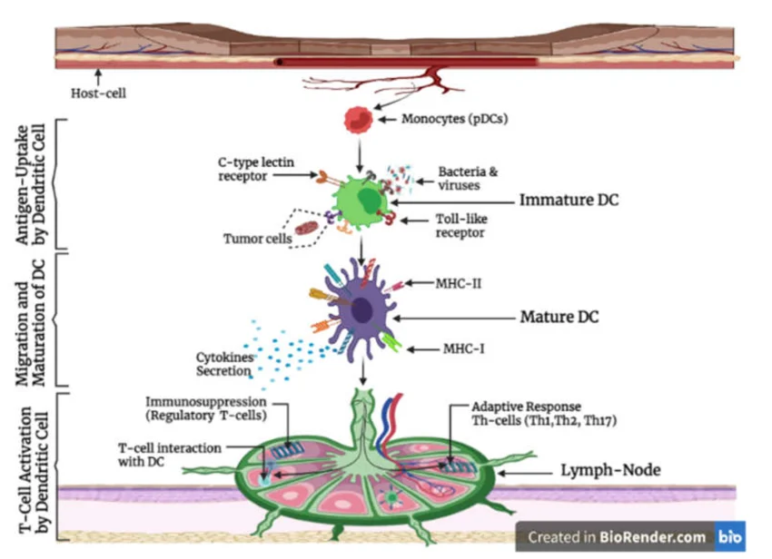
Dendritic cells (DCs) are essential antigen-presenting cells involved in tolerance induction.
Studies demonstrate that increasing dendritic cell populations enhances susceptibility to oral tolerance. DCs capture dietary antigens in the intestinal mucosa and transport them to lymphoid tissues where they interact with T lymphocytes.
This process controls immune activation and prevents excessive inflammatory responses against harmless food proteins.
Mesenteric Lymph Nodes as the Main Site of Oral Tolerance
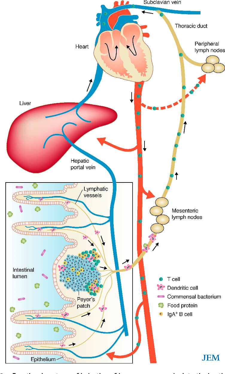
Mesenteric lymph nodes are specialized lymphoid structures located within the intestinal lymphatic network. They function as central regulators of mucosal immunity.
Research shows that oral tolerance is induced primarily in the MLNs rather than in Peyer’s patches or peripheral lymphoid organs.
Findings Supporting MLN Function
- Antigen presentation occurs predominantly in MLNs
- Mice lacking MLNs fail to develop oral tolerance
- Peyer’s patch deficiency alone does not abolish tolerance
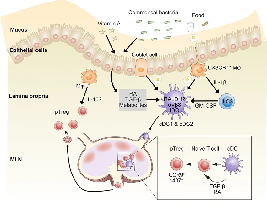
- Intestinal dendritic cells continuously migrate to MLNs carrying food antigens
These findings identify MLNs as the primary anatomical site where immune tolerance to dietary antigens is generated.
Migration of Antigen-Loaded Dendritic Cells
Intestinal dendritic cells sample luminal antigens from the gut epithelium and migrate through afferent lymphatic vessels to the MLNs.
Inside the MLNs, these antigen-loaded DCs:
- Present antigens to naïve T cells
- Induce T-cell proliferation under tolerogenic conditions
- Generate regulatory immune responses instead of inflammatory activation
The chemokine receptor CCR7 is critical for this migration process. Impaired CCR7 signaling prevents dendritic cell trafficking to MLNs and disrupts oral tolerance induction.
Local Versus Systemic Tolerance Induction
Although MLNs are the dominant site of oral tolerance induction, some evidence suggests that systemic lymphoid tissues may also contribute under certain conditions.
Food antigens entering the bloodstream through the hepatic portal circulation may reach:
- Peripheral lymph nodes
- Spleen
- Liver-associated immune tissues
Experimental studies show that intravenous or portal vein antigen exposure can also induce systemic immune tolerance.
However, under normal physiological conditions, MLNs remain the central and obligatory site for efficient oral tolerance generation.
Mesenteric Lymph Nodes and Commensal Bacteria Control
The intestine contains a massive population of commensal microorganisms that coexist with the host immune system.
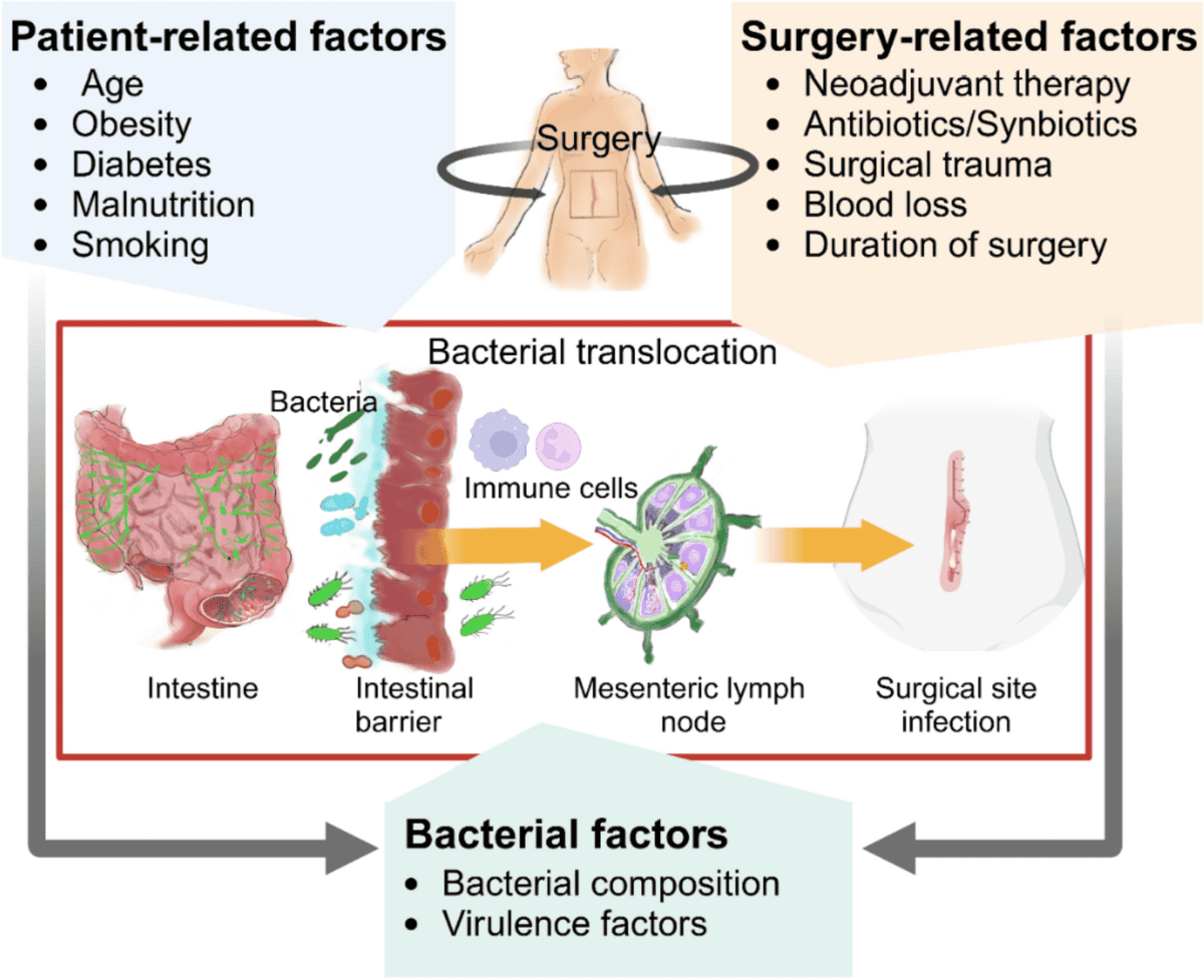
Unlike soluble food antigens, live bacteria are highly immunogenic. Despite continuous exposure to intestinal microbiota, the systemic immune system normally remains unresponsive to these organisms.
MLNs play a critical protective role by functioning as an immune “firewall.”
MLNs as an Immunological Firewall
Mesenteric lymph nodes prevent systemic dissemination of intestinal bacteria.
Protective Functions of MLNs
- Restrict migration of bacteria-loaded dendritic cells
- Prevent live commensal bacteria from entering systemic circulation
- Limit systemic immune activation against intestinal microbiota
In animals lacking MLNs, intestinal bacteria can reach the bloodstream and systemic lymphoid organs, leading to:
- Splenomegaly
- Lymphadenopathy
- Systemic immune activation
Thus, MLNs preserve immune compartmentalization between mucosal and systemic immunity.
IgA Production and Mucosal Immunity
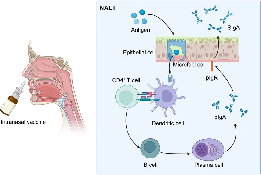
Intestinal dendritic cells also stimulate the production of IgA-producing B cells.
These B cells migrate through the lymphatic and circulatory systems to populate the intestinal lamina propria, where they differentiate into plasma cells that secrete IgA antibodies.
IgA plays a major role in:
- Neutralizing pathogens
- Regulating microbiota composition
- Protecting mucosal surfaces
Most IgA induction occurs in Peyer’s patches, while MLNs regulate microbial containment and immune tolerance.
Immune Communication Between Gut and Systemic Immunity
The intestinal immune system contains a large proportion of the body’s immune cells and antibody-producing plasma cells.
Mesenteric lymph nodes occupy a strategic position between:
- Mucosal immune tissues
- Systemic immune circulation
Because of this anatomical location, MLNs regulate:
- Antigen transport
- Immune tolerance
- Microbial containment
- Lymphocyte trafficking
- Adaptive immune activation
They function as a biological checkpoint controlling immune communication between the gut and the rest of the body.
Conclusion
Mesenteric lymph nodes are essential regulators of intestinal and systemic immune homeostasis. They serve as the primary site for oral tolerance induction by receiving antigen-loaded dendritic cells from the intestinal mucosa and promoting regulatory immune responses.
Key functions of MLNs include:
- Induction of tolerance to food antigens
- Regulation of CD4+ T-cell responses
- Prevention of food hypersensitivity
- Restriction of systemic bacterial dissemination
- Maintenance of immune compartmentalization
Overall, MLNs represent a critical anatomical and functional center of mucosal immunology, balancing immune tolerance, microbial control, and systemic immune protection.