

Introduction to Human Ocular Filariasis
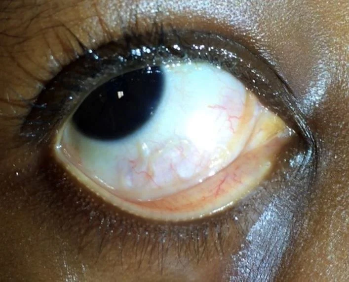
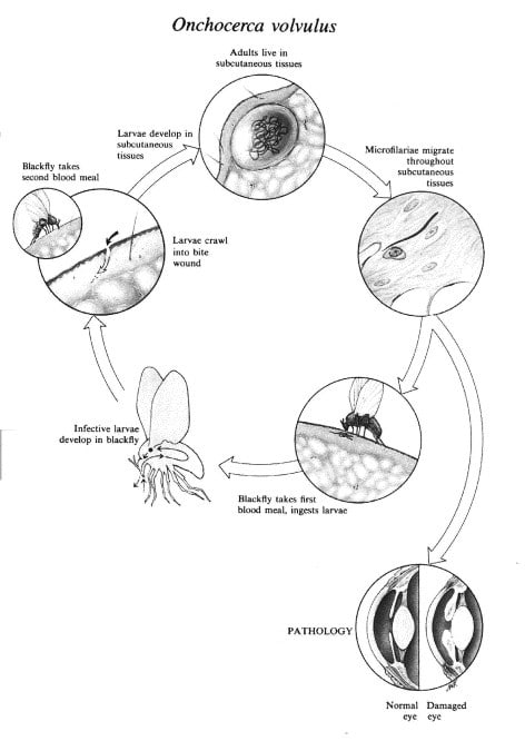
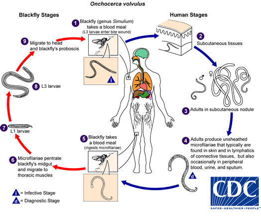
Helminth parasites remain an important cause of human ocular diseases in both developed and developing countries. Several parasitic nematodes are capable of invading ocular tissues and may lead to severe inflammation, visual impairment, or even blindness. One of the most well-known examples is river blindness caused by Onchocerciasis, a disease associated with millions of infections worldwide, particularly in Africa and parts of Latin America. In this condition, blindness mainly results from the host immune response against parasite microfilariae released into subcutaneous tissues.
Besides classical human filarial parasites such as Loa loa, Wuchereria bancrofti, and Brugia malayi, several parasites that normally infect domestic or wild animals may also act as zoonotic agents in humans. These include species of Dirofilaria, Onchocerca, Brugia, Molinema, and Pelecitus. Many of these parasites can invade ocular structures including the conjunctiva, eyelids, lacrimal glands, and even deeper ocular tissues.
Among emerging zoonotic filarial parasites, Onchocerca lupi has gained increasing attention because of its ability to infect both animals and humans, particularly at the ocular level.
Emergence of Onchocerca lupi as a Zoonotic Parasite
Onchocerca lupi was originally identified in periocular tissues of a wolf (Canis lupus) in Georgia. For many years, the parasite remained poorly understood, and its identification was initially questioned because Onchocerca species are more commonly associated with ungulates rather than canids. Later morphological and molecular investigations confirmed the validity of the species and corrected previous taxonomic uncertainties.
Subsequent veterinary studies demonstrated that O. lupi infects dogs in several European countries including Greece, Portugal, Hungary, Germany, and Switzerland, as well as in the United States. In canine hosts, the parasite produces ocular disease characterized by:
- Conjunctivitis
- Photophobia
- Excessive lacrimation
- Ocular discharge
- Exophthalmia
- Chronic periocular inflammation
The discovery of confirmed human infections strongly supports the zoonotic potential of this parasite.
Human Cases of Ocular Onchocerca lupi Infection
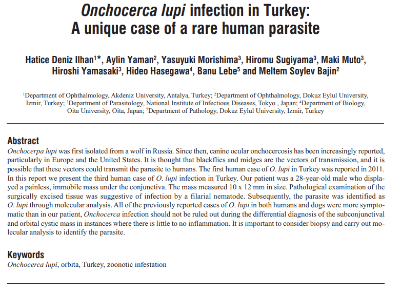
The study described two human ocular filariasis cases caused by Onchocerca lupi, one from Turkey and another from Tunisia. These findings provided additional evidence that humans can act as accidental hosts for this parasite.
Case from Turkey
A 26-year-old male patient from Istanbul presented with persistent irritation, redness, itching, and swelling of the right eye. Imaging revealed increased soft tissue density near the conjunctiva, initially suggesting inflammation or a foreign body. Standard treatment with corticosteroids and antibiotics failed to improve the condition.
During exploratory ocular surgery, clinicians discovered filamentous nematodes tightly attached beneath the subconjunctival tissue. The extracted worms measured approximately 10 cm in total length. Initially, the parasite was incorrectly suspected to be Dirofilaria repens.
Detailed histological and morphological analysis later confirmed that the nematode was an adult female Onchocerca lupi. Molecular testing based on mitochondrial markers further validated the diagnosis.
Case from Tunisia
The second case involved an 8-year-old child from the Kasserine region of Tunisia who experienced persistent pain in the right eye for several weeks. Examination revealed a subconjunctival mass that was surgically removed.
The parasite was originally misidentified as Dirofilaria immitis. However, later histopathological reassessment demonstrated features consistent with Onchocerca lupi. Unlike the Turkish case, surrounding host tissues were also available for microscopic evaluation, allowing a more detailed pathological examination.
This report represented one of the first confirmed indications of O. lupi infection in North Africa and highlighted the possibility that the parasite may circulate undetected in local animal populations.
Morphological Characteristics of Onchocerca lupi
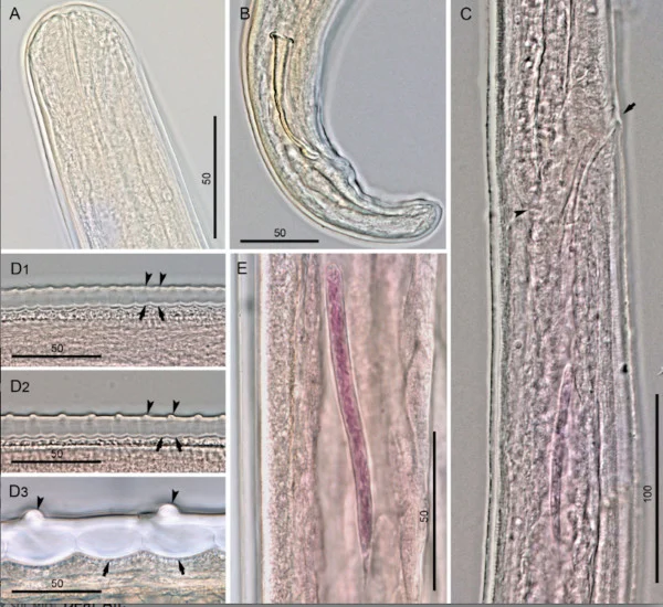
The diagnosis of O. lupi relies heavily on morphological and histological examination. Female worms possess distinctive cuticular structures that differentiate them from other filarial nematodes.
Key diagnostic features include:
- Thick multilayered cuticle
- External transverse ridges with rounded and undulated morphology
- Internal transverse striae
- Narrow lateral hypodermal cords
- Small buccal cavity
- Simple vagina without chambers or bends
- Coiled body structure within tissues
These features clearly distinguish O. lupi from Dirofilaria species:
- Dirofilaria repens has longitudinal cuticular ridges
- Dirofilaria immitis possesses a smooth cuticle without transverse ridging
The study emphasized that proper preservation of extracted nematodes is critical for accurate identification.
Molecular Diagnosis and DNA-Based Identification
Molecular analysis played a major role in confirming the identity of the parasite. Researchers amplified mitochondrial genes including:
- Cytochrome c oxidase subunit 1 (cox1)
- 12S ribosomal DNA
Sequence analysis demonstrated 99–100% similarity with previously reported O. lupi sequences available in GenBank databases.
Modern molecular tools combined with morphological examination provide highly reliable identification even when only small parasite fragments are available.
Zoonotic Transmission and Public Health Importance
The findings strongly support the zoonotic role of Onchocerca lupi. Dogs are currently considered the main reservoir hosts, although wild canids may also participate in transmission cycles.
Humans likely become infected through arthropod vectors that transmit infective larvae between hosts. However, the exact insect vector responsible for O. lupi transmission remains uncertain and requires additional investigation.
The study suggests that human infection may be underdiagnosed because:
- Cases are frequently misidentified as Dirofilaria infections
- Awareness among clinicians remains limited
- Routine molecular testing is rarely performed
- Epidemiological surveillance in animals is insufficient
Regions where infected dogs are present may therefore represent potential risk areas for human ocular infection.
Importance of Accurate Diagnosis
The study highlighted several important diagnostic recommendations for clinicians and ophthalmologists:
Proper Sample Collection
When parasites are surgically removed from ocular tissues:
- A portion should be preserved in ethanol for molecular testing
- Another portion can be processed for histology
Improper fixation may compromise DNA analysis and reduce diagnostic accuracy.
Differential Diagnosis
Onchocerca lupi should be considered in the differential diagnosis of subconjunctival or periocular nematode infections, particularly in endemic or suspected endemic regions.
Combined Diagnostic Approaches
The integration of:
- Histopathology
- Morphological examination
- DNA barcoding
- Molecular phylogenetics
provides the highest diagnostic reliability.
Clinical and Epidemiological Significance
This study demonstrated that Onchocerca lupi is not an isolated or accidental human parasite. Instead, it represents an emerging zoonotic filarial pathogen capable of causing ocular disease in humans across different geographic regions.
The parasite may circulate silently in canine populations while remaining undetected in humans until ocular symptoms appear. Because infections can mimic other ocular parasitic diseases, accurate identification is essential for epidemiological surveillance and patient management.
Future studies should focus on:
- Identifying insect vectors
- Understanding transmission dynamics
- Determining prevalence in dogs and wild animals
- Developing serological diagnostic tools
- Improving awareness among ophthalmologists and parasitologists
Conclusion
Human ocular filariasis caused by Onchocerca lupi represents an emerging zoonotic disease with increasing clinical importance. The parasite can infect subconjunctival and periocular tissues in humans and is frequently misdiagnosed as other filarial infections such as Dirofilaria species.
Morphological examination combined with molecular diagnostics has confirmed the zoonotic capability of O. lupi in both Europe and North Africa. Dogs are likely the primary reservoir hosts, although the complete transmission cycle remains incompletely understood.
Greater surveillance, improved diagnostic awareness, and further epidemiological studies are essential to better understand the global distribution and public health impact of this emerging ocular parasite.