

Introduction to Intestinal Immune Defense and Antigen Recognition
The gastrointestinal mucosa represents one of the largest and most complex immune interfaces in the body. Its primary physiological role is to facilitate the absorption of nutrients, vitamins, and digested food molecules while simultaneously protecting the host against harmful pathogens, toxins, and foreign antigens continuously entering through the digestive tract. Despite its selective barrier function, the intestinal epithelium is not completely impermeable. Various undigested macromolecules and antigenic substances can cross the mucosal surface and interact with the intestinal immune system.
Under healthy physiological conditions, exposure to dietary antigens generally promotes immune tolerance rather than inflammatory activation. This process is essential for maintaining intestinal homeostasis and preventing unnecessary immune reactions against harmless food proteins or commensal microbiota. However, when the intestinal barrier becomes damaged due to inflammation, infection, or chronic gastrointestinal disease, antigens may gain abnormal access to the gut-associated lymphoid tissue (GALT), bypassing normal regulatory pathways and triggering exaggerated immune responses.
Several inflammatory disorders, including celiac disease, ulcerative colitis, food hypersensitivity syndromes, and inflammatory bowel diseases, are associated with dysregulated mucosal immunity. Experimental evidence has shown that intestinal inflammation significantly increases the uptake and systemic penetration of undigested proteins and foreign antigens. Such alterations may disturb the balance between immune tolerance and hypersensitivity, potentially contributing to allergic and autoimmune intestinal diseases.
Understanding the mechanisms that regulate interactions between intestinal antigens and the mucosal immune system is therefore critical for improving knowledge of gastrointestinal immunopathology, oral tolerance, mucosal vaccination, and food allergy development.
Experimental Investigation of Intestinal Immune Responses
Objective of the Study
This study examined how different forms of intestinal mucosal damage influence local and systemic immune responses following antigen exposure. Researchers specifically evaluated whether antigens introduced through damaged intestinal tissues could stimulate stronger immune activation compared with antigens administered under normal physiological conditions.
The investigation focused on comparing immune reactions after antigen exposure through several experimental routes, including:
- Oral administration
- Direct delivery into Peyer’s patches
- Exposure through surgically traumatized intestinal mucosa
- Administration during chemically induced intestinal inflammation
The goal was to determine how mucosal integrity affects antigen uptake, antibody production, and activation of the gut-associated immune system.
Experimental Design and Animal Model
Laboratory Animals and Test Antigen
Male PVG strain rats weighing between 200 and 250 grams were used as the experimental model. Sheep red blood cells (SRBCs) served as the test antigen because they are widely used in immunological studies to evaluate antibody responses.
Before administration, SRBCs were purified and washed using phosphate-buffered saline (PBS) to remove contaminants and ensure experimental consistency.
Routes of Antigen Administration
Oral Antigen Exposure
One group of rats received SRBCs orally by gavage. This route simulated natural dietary antigen exposure occurring in the gastrointestinal tract under normal conditions.
Direct Peyer’s Patch Injection
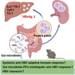
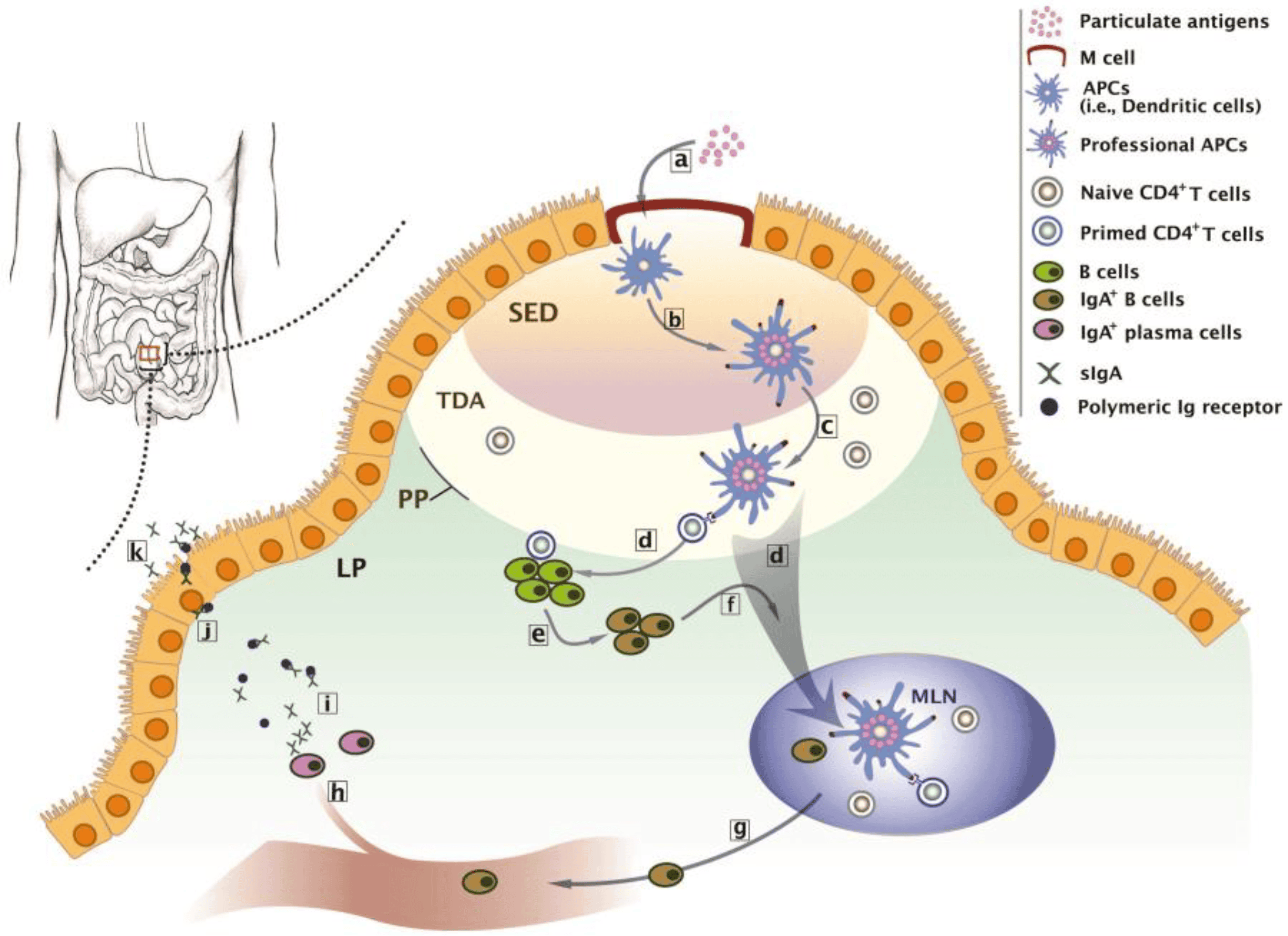
Another group received antigen injections directly into Peyer’s patches, which are specialized lymphoid structures located within the intestinal wall and play a central role in mucosal immune surveillance.
Antigen Delivery Through Traumatized Intestinal Tissue
To evaluate the effects of physical mucosal injury, researchers surgically exposed the ileum and gently damaged a small intestinal segment using a fine needle before applying the antigen directly onto the injured mucosal surface.
This model simulated conditions in which intestinal barrier disruption allows enhanced antigen penetration into immune tissues.
Chemically Induced Intestinal Inflammation
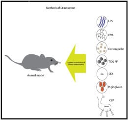
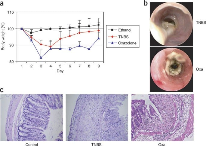
Additional experiments involved chronic administration of degraded iota carrageenan in drinking water to induce non-specific intestinal inflammation. Carrageenan exposure produced mucosal lesions and inflammatory alterations resembling pathological intestinal damage.
Following inflammation induction, animals were challenged orally with SRBCs to determine whether inflamed mucosa enhanced immune activation.
Assessment of Immune Responses
Serum and Biliary Antibody Analysis
Blood and bile samples were collected at multiple time points after antigen administration. Researchers measured anti-SRBC antibody production using standard haemagglutination assays.
The study evaluated both:
- Systemic immune responses through serum antibodies
- Local mucosal immunity through biliary antibody secretion
Detection of Antibodies Against Gut Microbiota
Researchers also assessed immune responses directed against intestinal bacteria using indirect immunofluorescence assays. Bacterial samples from rat gut flora cultures were exposed to serum and bile samples to determine levels of secretory IgA and other antibodies reactive against commensal microorganisms.
This analysis provided insight into how mucosal inflammation affects immune recognition of normal intestinal microbiota.
Findings of the Study
Minimal Immune Activation After Normal Oral Antigen Exposure
Rats receiving oral SRBCs under normal physiological conditions generated little or no detectable antibody response. This finding supports the concept of oral immune tolerance, in which the healthy gut suppresses excessive reactions against dietary antigens.
Strong Immune Responses After Direct GALT Exposure
Injection of SRBCs directly into Peyer’s patches produced rapid and substantial antibody production in both serum and bile. The biliary response appeared earlier and reached higher titres than the systemic serum response, demonstrating efficient activation of mucosal immunity.
These results confirm the central immunological role of Peyer’s patches in antigen recognition and mucosal antibody production.
Mucosal Injury Enhances Antigen Immunogenicity
Antigen application onto traumatized intestinal mucosa triggered significant systemic and local immune responses. Compared with intact mucosa, damaged tissues allowed increased antigen access to immune structures within the intestinal wall.
This observation suggests that disruption of epithelial integrity can transform normally tolerated antigens into highly immunogenic stimuli.
Carrageenan-Induced Inflammation Promotes Oral Immunization
Animals exposed to degraded carrageenan developed enhanced mucosal immune responses following oral antigen administration. Significant biliary antibody production against SRBCs was detected during active inflammation, whereas systemic serum antibodies remained limited.
Additionally, carrageenan-treated rats displayed markedly elevated antibody responses against normal gut bacteria, indicating increased immune reactivity toward intestinal microbiota during mucosal inflammation.
Interestingly, this effect was reversible. After discontinuation of carrageenan treatment and recovery of the intestinal mucosa, oral antigen exposure no longer induced strong antibody production.
Mechanisms Linking Mucosal Damage and Immune Activation
Increased Antigen Penetration Through Damaged Epithelium
Inflammatory lesions likely increased intestinal permeability, allowing antigens to penetrate deeper tissues and interact directly with GALT structures. Enhanced antigen translocation may therefore amplify immune activation and antibody synthesis.
Altered Macrophage Function
Carrageenan exposure may also modify macrophage activity and antigen processing pathways. Changes in macrophage-mediated immune regulation could contribute to abnormal antigen presentation and dysregulated immune responses.
These mechanisms may act simultaneously to disturb intestinal immune homeostasis.
Oral Tolerance and Intestinal Immune Regulation
Although transient mucosal antibody responses can occur after oral antigen exposure, prolonged exposure to many dietary proteins generally induces systemic immune unresponsiveness known as oral tolerance.
This tolerogenic mechanism is essential for preventing chronic intestinal inflammation and food hypersensitivity. Specialized regulatory pathways within the gut normally suppress harmful immune activation against harmless environmental antigens.
However, when mucosal integrity is compromised, these regulatory systems may fail. The resulting imbalance between tolerance and hypersensitivity could contribute to the pathogenesis of food allergies, inflammatory bowel disease, and other immune-mediated gastrointestinal disorders.
Conclusion
This study demonstrates that intestinal mucosal integrity plays a critical role in controlling both local and systemic immune responses to orally encountered antigens. Under healthy conditions, the gastrointestinal tract promotes immune tolerance and limits antibody production against dietary substances. In contrast, mucosal injury, inflammation, or direct antigen exposure to gut-associated lymphoid tissues can trigger strong immune activation and enhanced antibody responses.
The findings highlight the importance of epithelial barrier function in maintaining intestinal immune homeostasis and preventing pathological hypersensitivity reactions. These mechanisms are highly relevant to understanding food allergies, inflammatory bowel diseases, oral vaccine development, mucosal immunology, and antigen-specific immune regulation within the gastrointestinal tract.