


Introduction to Intestinal Epithelial Immunity
The intestinal epithelium is one of the most important defense systems in the human body. It forms a direct interface between the internal environment and the external world through continuous exposure to food, water, microorganisms, toxins, and environmental antigens. The gastrointestinal tract contains a massive epithelial surface area of nearly 200 m² in humans, making it one of the largest contact zones between host tissues and microbial communities.
This epithelial barrier is constantly exposed to bacteria, viruses, fungi, and parasites that enter the digestive tract. Some microorganisms are pathogenic and capable of causing infection, while others belong to the beneficial intestinal microbiota that support digestion, metabolism, immune development, and intestinal homeostasis. Because of this dual exposure, intestinal epithelial cells must maintain a delicate balance between immune defense and immune tolerance.
The intestinal microbiota consists of trillions of microorganisms that establish a symbiotic relationship with the host. These microbial populations contribute to nutrient metabolism, vitamin synthesis, immune maturation, and protection against pathogens. However, despite their beneficial role, intestinal microbes can become harmful if they cross the epithelial barrier or trigger uncontrolled inflammatory responses. Therefore, the intestinal epithelium acts as both a physical barrier and an active immune organ that regulates host–microbe interactions.
To maintain intestinal homeostasis, epithelial cells have evolved multiple defense strategies, including mucus secretion, antimicrobial peptide production, microbial sensing systems, autophagy activation, cytokine secretion, and regulation of adaptive immunity. These mechanisms protect host tissues from microbial invasion while preserving beneficial microbial communities within the gut lumen.
The Intestinal Microbiota and Its Biological Importance
Composition of the Gut Microbiota
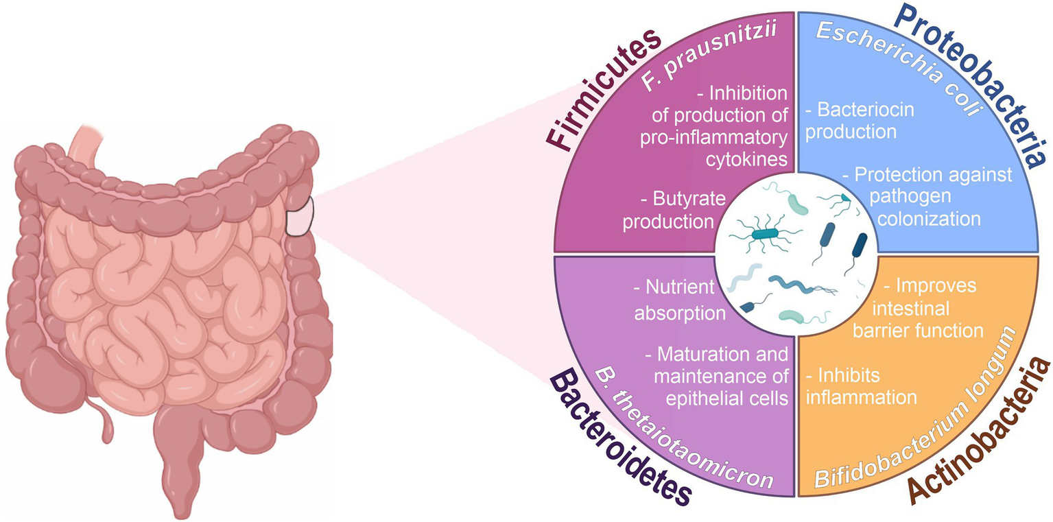
The human intestinal microbiota contains hundreds to thousands of bacterial species. Molecular sequencing technologies have shown that the dominant bacterial groups belong mainly to two phyla:
- Firmicutes
- Bacteroidetes
Additional bacterial populations include members of:
- Proteobacteria
- Actinobacteria
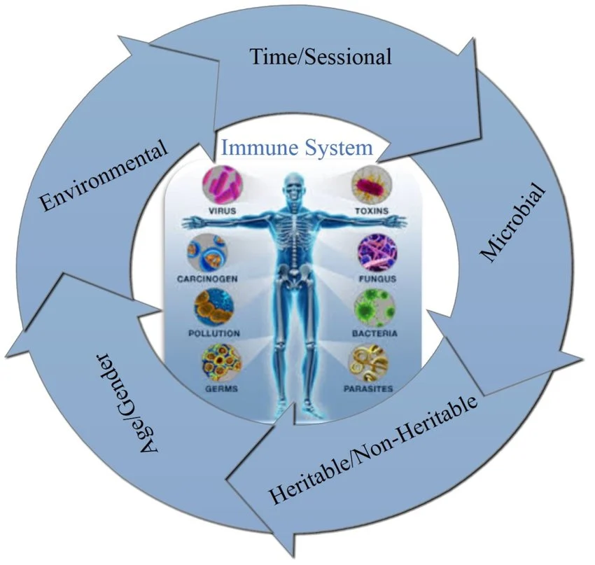
The composition of the microbiota varies according to:
- Diet
- Age
- Lifestyle
- Environmental exposure
- Antibiotic use
- Host genetics
Important bacterial species found in the intestine include:
- Bacteroides thetaiotaomicron
- Bacteroides fragilis
- Lactobacillus species
- Clostridia species
- Enterococcus species
These microbial communities form highly dynamic ecosystems that continuously interact with intestinal epithelial cells.
Functional Roles of the Intestinal Microbiota
The gut microbiota performs numerous physiological functions that are essential for human health.
Enhancement of Nutrient Digestion
Many dietary compounds, particularly complex polysaccharides from plants, cannot be digested directly by human enzymes. Intestinal bacteria produce specialized enzymes such as glycosyl hydrolases that degrade these complex carbohydrates into absorbable nutrients.
This microbial metabolism:
- Increases caloric extraction from food
- Produces short-chain fatty acids
- Supports intestinal epithelial nutrition
- Regulates energy metabolism
Regulation of Immune System Development
The microbiota plays a central role in shaping the immune system. Intestinal bacteria regulate the differentiation and activation of multiple immune cell populations, including:
- T helper 17 (TH17) cells
- Regulatory T cells (Tregs)
- B lymphocytes
- Dendritic cells
Microbial signals also influence the balance between pro-inflammatory and anti-inflammatory immune responses, helping maintain mucosal immune tolerance.
Effects on Metabolism and Neurological Function
Emerging research demonstrates that gut microbes influence:
- Fat storage
- Glucose metabolism
- Hormonal signaling
- Brain development
- Nervous system communication
Alterations in microbiota composition have been associated with inflammatory bowel disease, obesity, metabolic syndrome, and neurodevelopmental disorders.
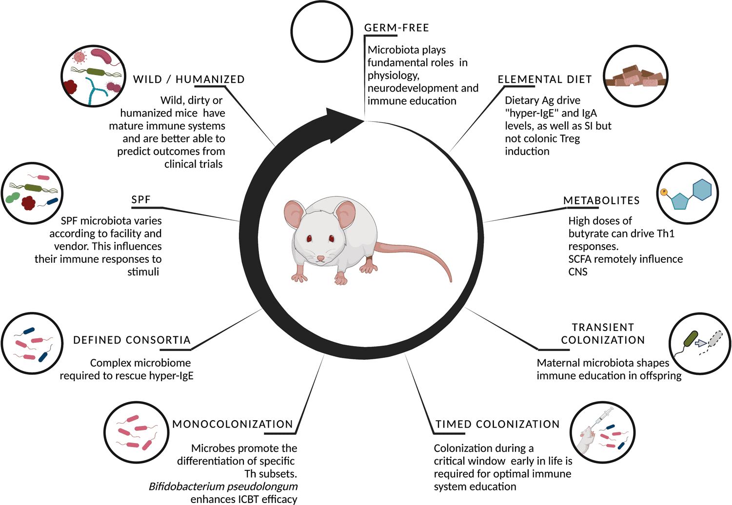
Germ-Free Animal Models in Intestinal Immunology
Germ-free animals are raised in sterile environments without exposure to microorganisms. These models are essential for studying epithelial immunity and microbiota-host interactions.
Comparisons between germ-free and colonized animals have demonstrated that intestinal microbes regulate:
- Antimicrobial protein expression
- Autophagy activation
- Immune cell differentiation
- Cytokine production
- Barrier maturation
Gnotobiotic models, where defined microbial populations are introduced into sterile animals, provide powerful tools for understanding microbial contributions to intestinal immunity.
Cellular Components of the Intestinal Epithelial Barrier
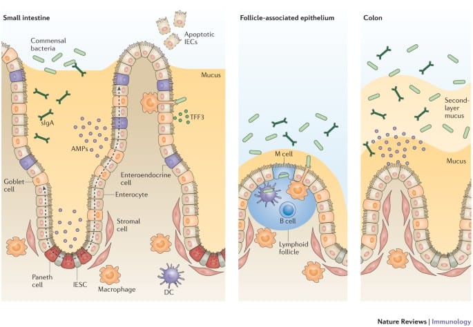
The intestinal epithelial barrier is composed of several specialized cell lineages that contribute to mucosal defense and immune regulation.
Enterocytes
Enterocytes are the most abundant epithelial cells in the intestine. They create a strong physical barrier through tight junctions and actively participate in immune defense.
Major enterocyte functions include:
- Production of antimicrobial proteins
- Activation of autophagy pathways
- Cytokine secretion
- Transport of immunoglobulin A (IgA)
Enterocytes also coordinate communication between luminal microbes and underlying immune cells.
Goblet Cells
Goblet cells are specialized secretory epithelial cells responsible for mucus production.
Functions of Goblet Cells
Mucus Secretion
Goblet cells produce mucins that form a gel-like mucus layer covering the intestinal surface. This mucus layer:
- Prevents bacterial contact with epithelial cells
- Limits pathogen invasion
- Protects tissues from mechanical damage
Immune Regulation
Goblet cells also secrete:
- Trefoil factors
- Resistin-like molecules
- Immunomodulatory proteins
Recent studies show that goblet cells participate in antigen sampling and antigen delivery to dendritic cells.
Paneth Cells
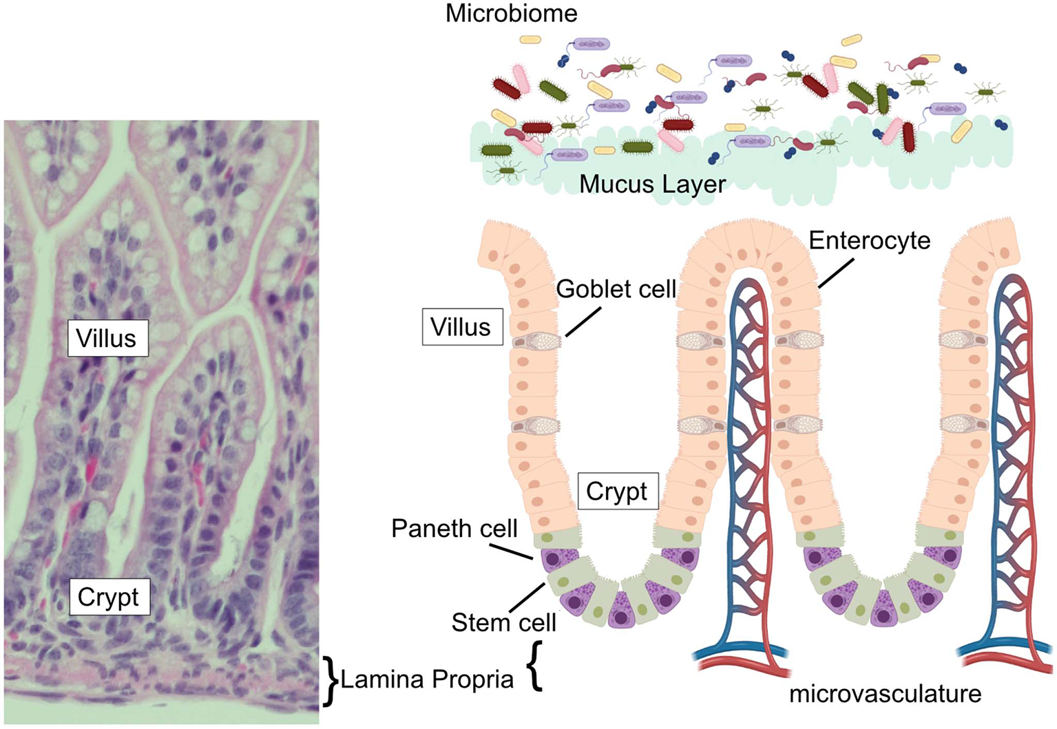
Paneth cells are located at the base of small intestinal crypts and are major producers of antimicrobial proteins.
Paneth Cell Secretory Products
Paneth cells release:
- α-defensins
- Lysozyme
- Phospholipase A2
- C-type lectins
- Antimicrobial peptides
These molecules directly destroy bacteria and regulate microbial populations near epithelial surfaces.
Paneth cells also support intestinal stem cells by secreting:
- EGF
- WNT3
- Notch ligands
This makes them essential for epithelial regeneration and tissue renewal.
Enteroendocrine Cells
Enteroendocrine cells function as hormone-producing epithelial cells that regulate digestion and metabolism.
They secrete molecules such as:
- Glucagon-like peptide
- Gastric inhibitory peptide
- Vasoactive intestinal peptide
These hormones control:
- Nutrient absorption
- Gut motility
- Appetite regulation
- Metabolic homeostasis
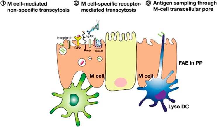
M Cells and Antigen Sampling
Microfold cells (M cells) are specialized epithelial cells involved in antigen transport.
M cells:
- Capture luminal antigens
- Deliver microorganisms to immune tissues
- Activate adaptive immune responses
Although essential for mucosal immunity, M cells may also serve as entry points for invasive pathogens.
Pattern Recognition Receptors in Epithelial Immunity
Intestinal epithelial cells recognize microorganisms using pattern recognition receptors (PRRs).
Toll-Like Receptors (TLRs)
TLRs detect conserved microbial molecules including:
- Lipopolysaccharide (LPS)
- Flagellin
- Peptidoglycan
- Bacterial DNA
Activation of TLR signaling stimulates inflammatory pathways through NF-κB activation and cytokine production.
Important epithelial TLRs include:
- TLR2
- TLR4
- TLR5
- TLR9
These receptors regulate antimicrobial defense and epithelial immune responses.
NOD-Like Receptors and Inflammasomes
NOD-like receptors (NLRs) are intracellular microbial sensors.
NOD1 and NOD2
NOD receptors recognize bacterial peptidoglycan fragments and activate inflammatory signaling pathways.
NOD2 is strongly associated with Crohn’s disease susceptibility and regulates:
- Defensin production
- Paneth cell activity
- Microbiota composition
Inflammasomes
Inflammasomes are multiprotein complexes that regulate inflammatory cytokine activation.
Important inflammasome components include:
- NLRP6
- ASC
- Caspase-1
Inflammasomes control:
- IL-1β production
- IL-18 secretion
- Mucus secretion
- Barrier integrity
Defective inflammasome signaling contributes to intestinal inflammation and dysbiosis.
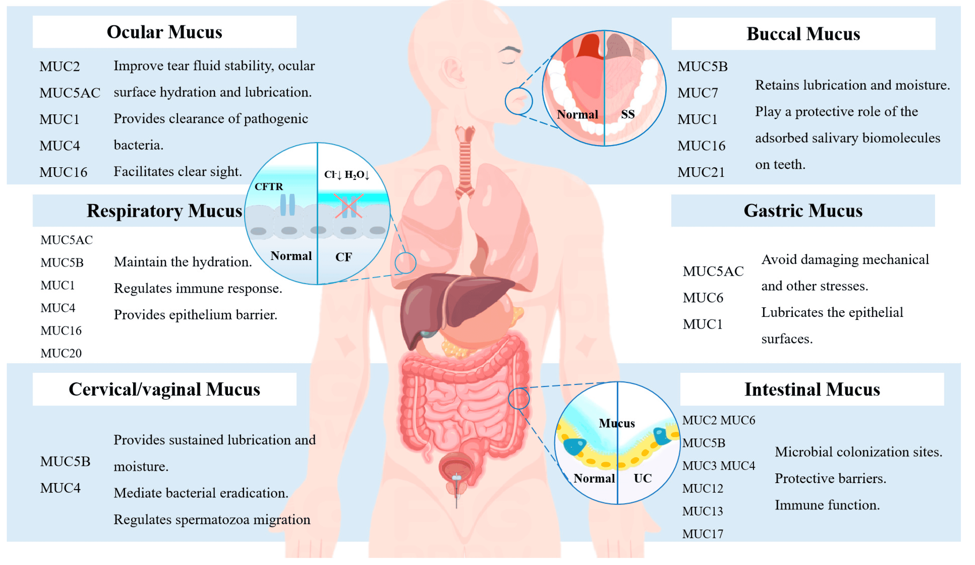
The Intestinal Mucus Barrier
The mucus layer is a critical component of epithelial defense.
Structure of the Mucus Layer
The intestinal mucus barrier contains:
- Outer mucus layer
- Colonized by bacteria
- Inner mucus layer
- Resistant to bacterial penetration
Goblet cell-derived mucins form the structural framework of this protective barrier.

Functions of Intestinal Mucus
The mucus layer:
- Prevents bacterial invasion
- Concentrates antimicrobial proteins
- Protects epithelial cells
- Maintains host–microbiota separation
Defects in mucus production are strongly linked to inflammatory bowel disease.
Intestinal Antimicrobial Proteins
Intestinal epithelial cells produce diverse antimicrobial proteins that directly eliminate microbes.
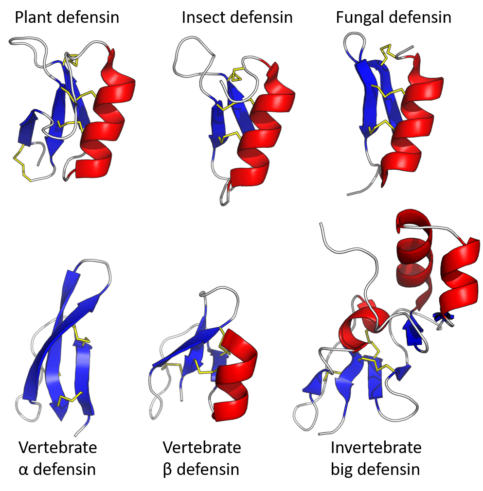
Defensins
Defensins are small cationic peptides that disrupt bacterial membranes.
Major defensin classes include:
- α-defensins
- β-defensins
- θ-defensins
These molecules exhibit broad antimicrobial activity against:
- Gram-positive bacteria
- Gram-negative bacteria
- Fungi
- Viruses
Paneth cells are major producers of α-defensins.
Lectins
C-type lectins such as RegIIIγ recognize bacterial cell wall structures and kill microbes through membrane disruption.
RegIII proteins are particularly effective against Gram-positive bacteria and help maintain spatial separation between microbes and epithelial cells.
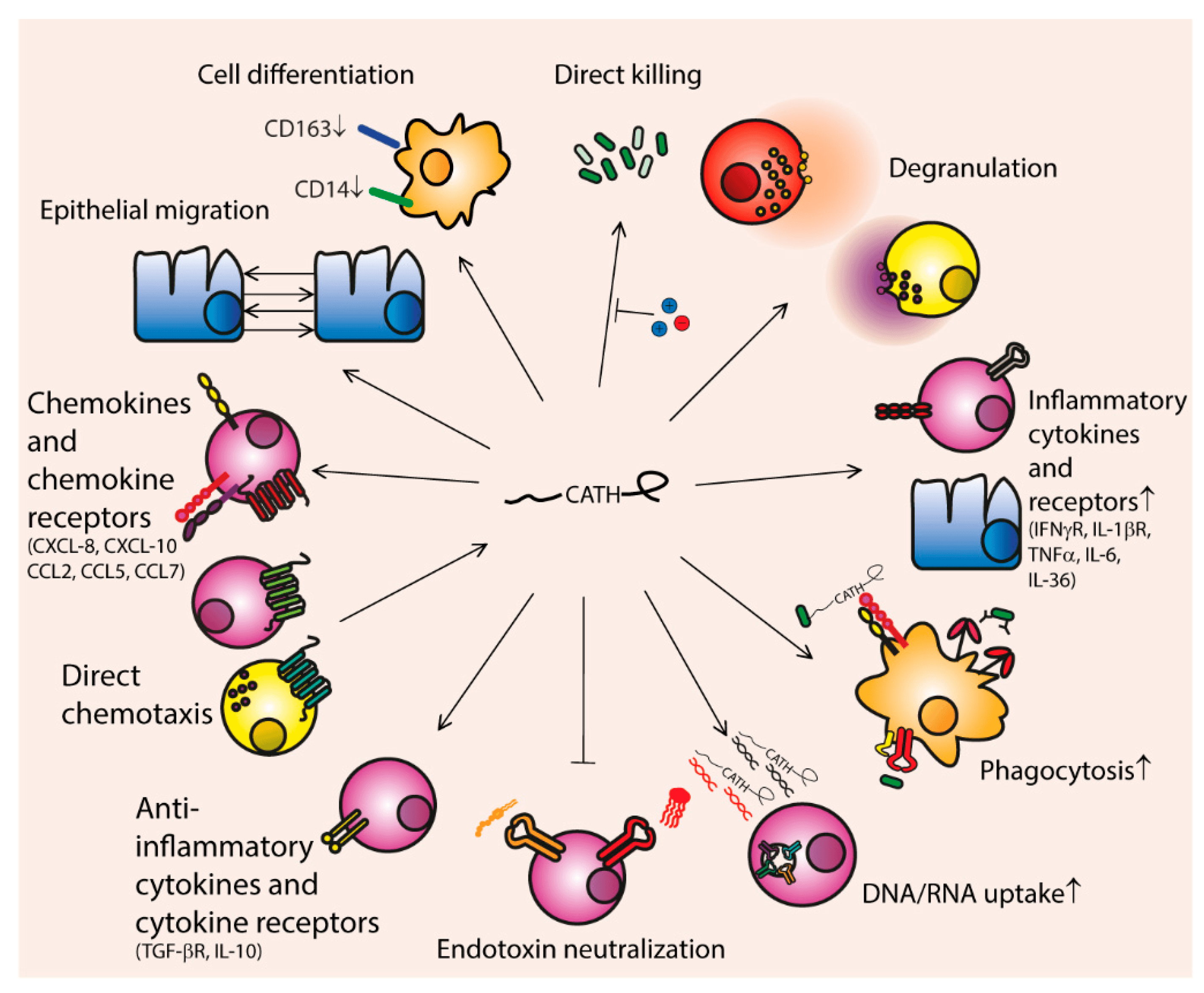
Cathelicidins
Cathelicidins are membrane-active antimicrobial peptides with broad-spectrum antimicrobial activity.
Human LL-37 is an important cathelicidin involved in:
- Bacterial killing
- Immune cell recruitment
- Cytokine regulation
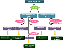
Lysozyme and Phospholipase A2
Lysozyme degrades bacterial peptidoglycan, while phospholipase A2 hydrolyzes bacterial membrane phospholipids.
Both proteins are highly expressed in Paneth cells and contribute to mucosal antimicrobial defense.
Lipocalins and Nutritional Immunity
Lipocalins inhibit bacterial growth by sequestering iron-containing siderophores.
This mechanism deprives bacteria of essential nutrients and limits microbial proliferation.
Regulation of Antimicrobial Protein Expression
Antimicrobial proteins are tightly regulated through:
- TLR signaling
- NOD signaling
- Cytokine pathways
- Microbial stimulation
- Developmental regulation
Some antimicrobial proteins are constitutively expressed, while others require microbial activation.
Autophagy in Intestinal Epithelial Cells
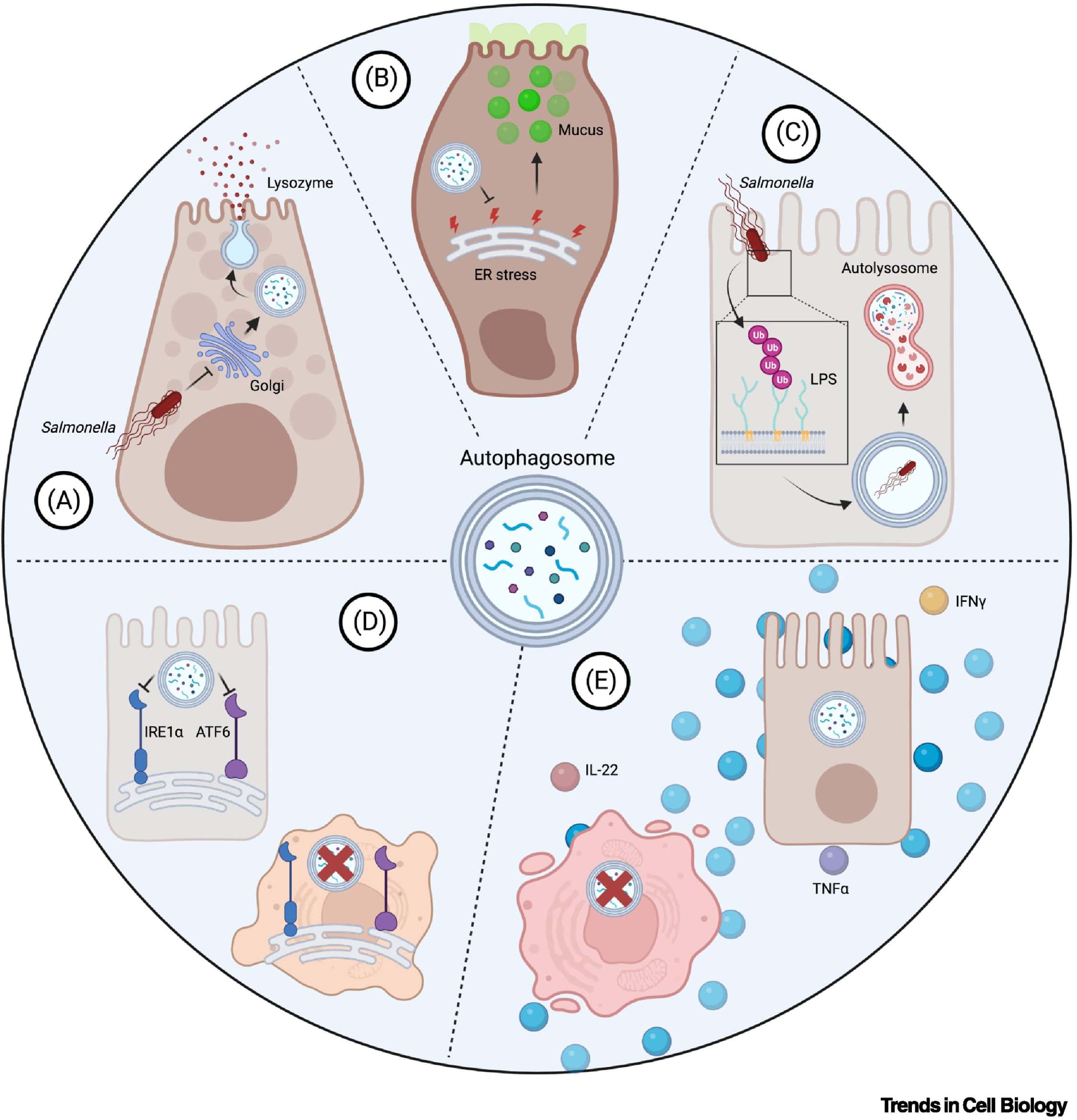
Autophagy is a cellular degradation pathway that protects epithelial cells against intracellular pathogens.
Functions of Epithelial Autophagy
Autophagy:
- Eliminates invasive bacteria
- Maintains cellular homeostasis
- Regulates Paneth cell granule secretion
- Controls goblet cell mucus release
Key autophagy proteins include:
- ATG5
- ATG16L1
- ATG7
Mutations in autophagy genes are strongly associated with Crohn’s disease.
Epithelial Regulation of Adaptive Immunity
Intestinal epithelial cells actively regulate adaptive immune responses.
Immunoglobulin A (IgA) Transport
Epithelial cells transport IgA across the mucosal barrier through the polymeric immunoglobulin receptor (pIgR).
Secretory IgA:
- Neutralizes pathogens
- Prevents microbial invasion
- Maintains microbial compartmentalization
Cytokine Production
Epithelial cells produce multiple cytokines involved in immune regulation, including:
- APRIL
- BAFF
- TSLP
- IL-22-responsive factors
These cytokines regulate:
- B-cell maturation
- IgA production
- Immune tolerance
- Anti-inflammatory responses
Antigen Presentation and Immune Sampling
Epithelial cells participate in antigen delivery to immune cells.
Important mechanisms include:
- M cell-mediated antigen transport
- Goblet cell antigen delivery
- Dendritic cell sampling
- CD1d-mediated lipid antigen presentation
These pathways coordinate mucosal immune surveillance and adaptive immunity.
Clinical Relevance of Intestinal Epithelial Immunity
Disruption of epithelial immune mechanisms contributes to numerous intestinal diseases, including:
- Crohn’s disease
- Ulcerative colitis
- Colorectal cancer
- Chronic inflammation
- Dysbiosis
- Infectious enteritis
Defects in:
- Mucus production
- Autophagy
- Antimicrobial peptide secretion
- PRR signaling
- Barrier integrity
can all promote intestinal pathology.
Future Perspectives in Epithelial Immunology
Research in epithelial immunology continues to reveal the complex interactions between epithelial cells and microbial ecosystems.
Future studies are expected to focus on:
- Host–microbiota signaling pathways
- Virome and fungal interactions
- Personalized microbiome therapies
- Barrier-targeted therapeutics
- Immune modulation strategies
- Probiotic and postbiotic interventions
Understanding epithelial immune regulation may lead to new therapeutic approaches for inflammatory, infectious, metabolic, and autoimmune diseases.
Conclusion
The intestinal epithelium is far more than a passive physical barrier. It is a highly dynamic and immunologically active tissue that continuously monitors microbial signals, regulates inflammation, produces antimicrobial defenses, and coordinates adaptive immune responses.
Through mucus production, antimicrobial protein secretion, microbial sensing pathways, autophagy activation, and immune communication, epithelial cells maintain a balanced relationship with the intestinal microbiota while protecting host tissues from infection and inflammation.
Disruption of these epithelial defense systems contributes directly to intestinal disease pathogenesis. Therefore, understanding epithelial cell contributions to intestinal immunity is essential for advancing modern gastroenterology, microbiology, mucosal immunology, and microbiome-based therapeutics.