

Introduction to Intestinal Immunity
The intestinal immune system represents one of the most specialized and highly organized defense networks in the human body. Most antigens, microorganisms, dietary proteins, and environmental particles enter the body through mucosal surfaces such as the gastrointestinal tract and respiratory system. Because of this constant exposure, mucosal tissues have evolved a unique immune architecture that differs significantly from systemic immunity.
Unlike the systemic immune system, mucosal immunity must maintain a delicate balance between protection and tolerance. It must rapidly eliminate harmful pathogens while simultaneously avoiding excessive inflammatory reactions against harmless food antigens, commensal microbiota, and environmental molecules. This dual function makes intestinal immunity one of the most complex components of human immunology.
The mucosal immune system includes immune tissues associated with the gastrointestinal tract, respiratory tract, and urogenital tract, as well as exocrine glands such as salivary glands, mammary glands, pancreatic tissue, and lacrimal glands. Among these compartments, the intestinal mucosa contains the largest concentration of immune cells in the body.
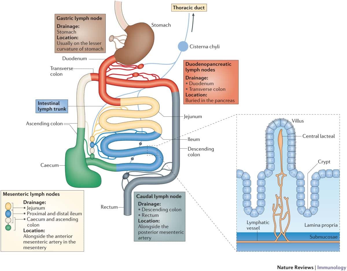
Structural Organization of the Mucosal Immune System
Large Surface Area and Continuous Antigen Exposure
The gastrointestinal tract possesses an extremely large functional surface area designed for nutrient absorption and digestion. In humans, the intestinal mucosal surface covers approximately 400 square meters, which is nearly 200 times larger than the skin surface.
This enormous interface is continuously exposed to:
- Dietary antigens
- Commensal bacteria
- Viruses
- Fungi
- Parasites
- Environmental toxins
- Pathogenic microorganisms
Every meal and every breath introduce foreign antigens into mucosal tissues. Consequently, the intestinal immune system must constantly monitor and regulate immune responses without damaging the epithelial barrier that is essential for life.
Because of this persistent antigenic stimulation, mucosal tissues contain the highest concentration of lymphocytes in the body. In fact, intraepithelial T cells located in the small intestine may account for nearly 60% of all T lymphocytes present in humans.
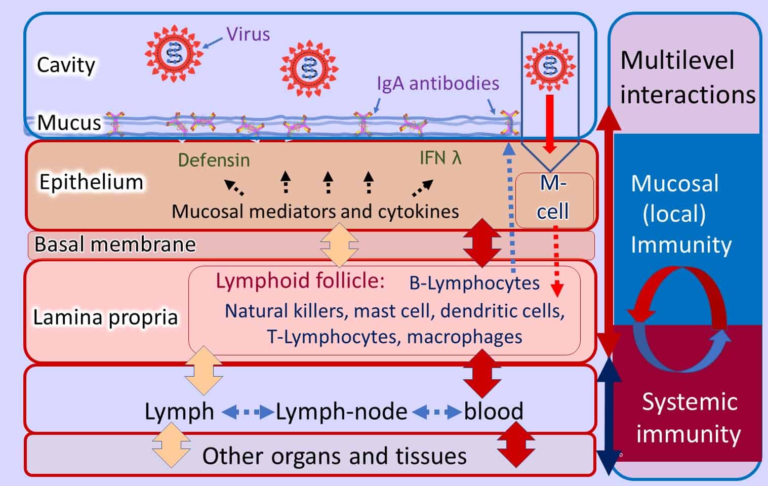
Gut-Associated Lymphoid Tissue (GALT)
Components of GALT
The gut-associated lymphoid tissue (GALT) forms the central immune compartment of the intestine. It includes:
- Peyer’s patches
- Mesenteric lymph nodes
- Lamina propria lymphocytes
- Intraepithelial lymphocytes
These structures function together to initiate, regulate, and distribute mucosal immune responses.
Peyer’s Patches
Peyer’s patches are specialized lymphoid follicles located beneath the intestinal epithelium. They resemble secondary lymphoid organs and contain organized T-cell and B-cell zones.
A distinctive feature of Peyer’s patches is their close association with the follicle-associated epithelium (FAE), which contains specialized microfold cells known as M cells.
Functions of M Cells
M cells play a critical role in mucosal antigen sampling by:
- Capturing luminal antigens
- Transporting microorganisms
- Delivering antigens to antigen-presenting cells
- Initiating adaptive immune responses
Many invasive pathogens, including Salmonella, Yersinia, and Shigella, exploit M-cell transport pathways to invade intestinal tissues.
Intestinal Epithelial Barrier and Immune Crosstalk
Epithelial Cells as Immune Regulators
Intestinal epithelial cells are not passive physical barriers. They actively participate in immune regulation through cytokine secretion and communication with immune cells.
Enterocytes produce numerous immunomodulatory molecules, including:
- IL-1
- IL-6
- IL-7
- IL-8
- TGF-β
- MIP-1β
These cytokines regulate:
- Lymphocyte activation
- Epithelial repair
- Barrier integrity
- Inflammatory signaling
- Cell differentiation
Conversely, cytokines secreted by immune cells influence epithelial function, permeability, ion transport, and MHC expression.
This bidirectional communication forms a dynamic epithelial–immune network essential for intestinal homeostasis.
Lymphocyte Trafficking and Mucosal Homing
Selective Migration of Immune Cells
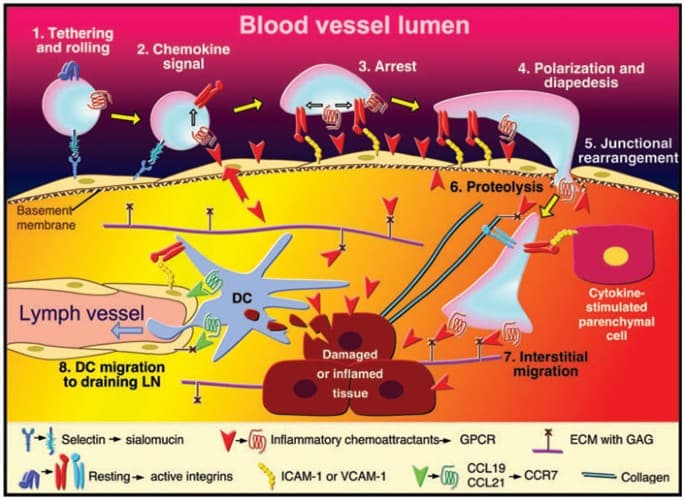
Activated lymphocytes generated within mucosal tissues preferentially migrate back to mucosal surfaces. This selective recirculation depends on specialized adhesion molecules and integrins.
The most important molecule involved is:
α4β7
This integrin interacts with mucosal vascular adhesion molecules such as MAdCAM-1.
Functions of α4β7 Integrin
The α4β7 integrin enables:
- Lymphocyte homing to intestinal tissues
- Retention within mucosal compartments
- Migration across endothelial barriers
- Maintenance of localized immune responses
Disruption of α4β7-mediated trafficking severely impairs intestinal immune surveillance.
Antigen Presentation in the Intestine
Multiple Antigen-Presenting Cell Types
The intestinal mucosa contains several antigen-presenting cell (APC) populations, including:
- Dendritic cells
- Macrophages
- Activated B cells
- Intestinal epithelial cells
Each APC type may influence immune outcomes differently, determining whether tolerance or active immunity develops.
Dendritic Cells and Oral Antigen Sampling
Dendritic cells located within Peyer’s patches and the lamina propria capture luminal antigens and migrate to mesenteric lymph nodes where they activate naïve T cells.
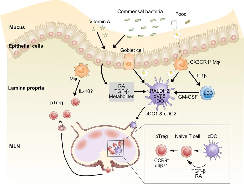
These dendritic cells can promote:
- Protective immunity
- Regulatory T-cell differentiation
- Oral tolerance
- Cytokine-mediated suppression
The final outcome depends heavily on the inflammatory context and co-stimulatory signals present during antigen presentation.
Oral Tolerance and Immune Regulation
Mechanisms of Oral Tolerance
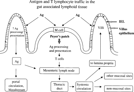
One of the most important features of intestinal immunity is oral tolerance, a process in which orally administered antigens induce systemic immune unresponsiveness.
This mechanism prevents harmful immune reactions against:
- Dietary proteins
- Commensal microbiota
- Environmental antigens
Oral tolerance involves several immunological processes:
- T-cell anergy
- Clonal deletion
- Regulatory T-cell activation
- Anti-inflammatory cytokine production
Key regulatory cytokines include:
- IL-10
- TGF-β
- IL-4
Intraepithelial Lymphocytes (IELs)
Specialized Immune Cells of the Epithelium
Intraepithelial lymphocytes are unique T cells embedded directly between intestinal epithelial cells.
Approximately 80–90% of IELs are CD8-positive T cells, many of which express unusual receptors and activation pathways distinct from peripheral T lymphocytes.
These cells participate in:
- Epithelial surveillance
- Cytotoxic defense
- Barrier maintenance
- Tissue repair
- Regulation of inflammation
IELs are positioned extremely close to luminal antigens, making them the first immune cells encountered by invading pathogens.
Gamma Delta (γδ) T Cells in Intestinal Immunity
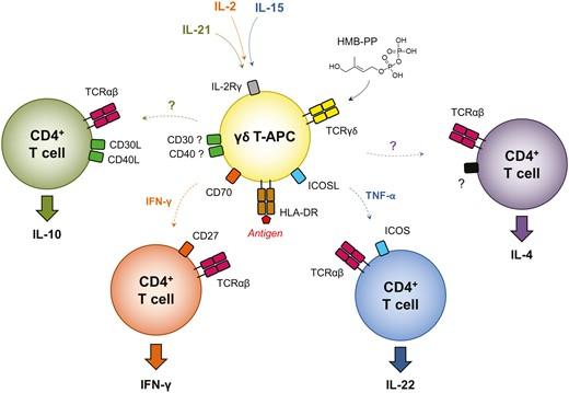
A significant proportion of IELs express gamma delta T-cell receptors rather than conventional alpha beta TCRs.
γδ T cells
These γδ T cells contribute to:
- Rapid antimicrobial defense
- Epithelial repair
- Cytokine secretion
- Immune regulation
- Maintenance of barrier integrity
Unlike classical T cells, many γδ IELs display limited antigen specificity and may recognize stress-induced epithelial molecules instead of conventional peptide antigens.
Extrathymic T-Cell Development in the Intestine
Intestinal Epithelium as a Lymphoid Organ
Research suggests that the intestinal mucosa may function as an alternative site of T-cell differentiation independent of the thymus.
Evidence supporting extrathymic lymphopoiesis includes:
- Presence of immature lymphoid precursors
- Expression of recombination genes
- Development of unique IEL populations
- Specialized epithelial microenvironments
This discovery has major implications for understanding immune system development and mucosal immunobiology.
Mucosal Immunopathology and Inflammatory Disease
Breakdown of Immune Regulation
Failure to properly regulate mucosal immune responses can result in chronic inflammatory disorders such as:
- Crohn’s disease
- Ulcerative colitis
- Celiac disease
- Asthma
- Allergic inflammatory disorders
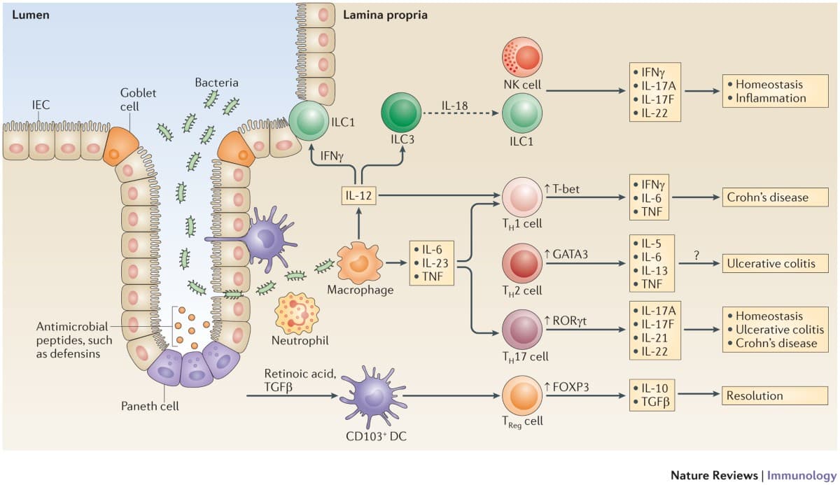
These diseases are often associated with excessive activation of mucosal CD4+ T cells and abnormal cytokine production.
Cytokines in Intestinal Inflammation
Several pro-inflammatory cytokines contribute directly to intestinal pathology, including:
- TNF-α
- IFN-γ
- IL-12
- IL-1
- TNF-β
These cytokines disrupt epithelial barrier integrity, stimulate inflammatory cell recruitment, and promote tissue injury.
Among them, TNF-α plays a major pathogenic role in inflammatory bowel disease.
TNF-αTNF
Modern biologic therapies targeting TNF-α have significantly improved treatment outcomes in Crohn’s disease and ulcerative colitis.
Clinical Importance of Mucosal Immunity
Understanding intestinal immunity has major implications for:
- Vaccine development
- Autoimmune disease therapy
- Inflammatory bowel disease treatment
- Oral immunotherapy
- Microbiome-based therapies
- Cancer immunology
Mucosal vaccine strategies are particularly important because immune responses induced directly at mucosal surfaces generate stronger local protection than systemic vaccination alone.
Conclusion
The intestinal immune system is a highly specialized and tightly regulated immune network designed to maintain the balance between defense and tolerance. Through the coordinated activity of Peyer’s patches, dendritic cells, epithelial cells, intraepithelial lymphocytes, cytokines, and mucosal adhesion molecules, the gut immune system protects the host from pathogens while preserving tolerance to harmless antigens and commensal microorganisms.
Disruption of this delicate equilibrium can lead to chronic inflammatory diseases such as Crohn’s disease, ulcerative colitis, and celiac disease. Continued research into mucosal immunology is essential for advancing vaccine technology, immunotherapy, microbiome research, and the treatment of autoimmune and inflammatory disorders.